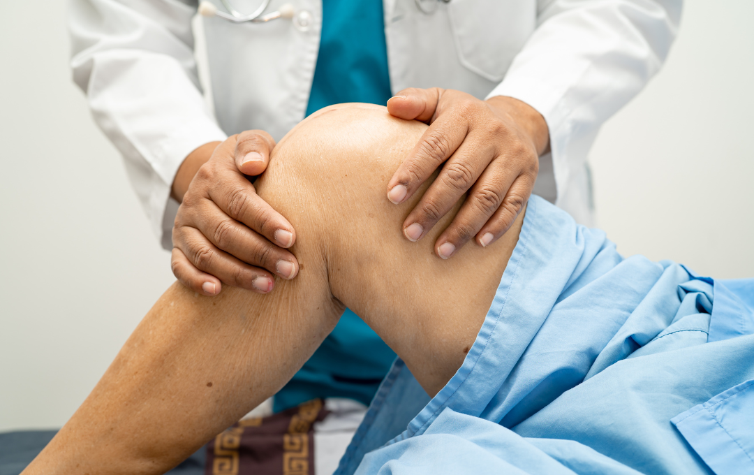
Imagine that one day, your knee starts to give out. It hurts when you move around, and it seems swollen compared to your other knee. You might have shrugged it off and iced it, thinking that it was just a minor injury that would resolve itself you’re still dealing with the same knee pain and swelling, except it seems to be getting worse. You visit your health care provider, who orders medical imaging tests to determine if you have knee arthritis.
Knee arthritis can be more than just knee pain and swelling. This degenerative disease can significantly impact mobility, making it harder to complete daily tasks as cartilage in the knee erodes. While X-rays can show bone damage from knee arthritis, they are not enough for a complete diagnosis. A specialist must combine the X-ray results with your physical symptoms and other tests to determine the best treatment plan.
Various medical imaging tools play a crucial role in diagnosing knee arthritis and guiding treatment options, such as genicular artery embolization (GAE). Below, we will discuss how X-rays help in this process, what their limitations are, and how other diagnostic tools can diagnose and treat knee arthritis.
Do You Really Need an X-Ray for Knee Arthritis Symptoms?
If you’re dealing with knee pain, stiffness, swelling, or trouble moving the joint, then yes – an X-ray is often the first step in understanding what’s going on. Many people with knee arthritis experience symptoms such as:
-
Persistent or worsening knee pain
-
Stiffness after sitting or resting
-
Swelling or inflammation around the joint
-
A grinding or cracking sensation (crepitus)
-
Pain that worsens with activity
-
Reduced range of motion
-
Difficulty climbing stairs or standing up
-
Knee instability or feeling like the knee might “give out”
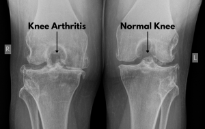
Most knee arthritis diagnoses begin with a simple X-ray. This imaging test shows joint space narrowing, bone spurs, and structural changes that confirm osteoarthritis. While an X-ray is a start, your doctor may order an MRI to evaluate cartilage loss or soft tissue damage if your symptoms are more complex.
Learn More About Knee Arthritis
How X-Rays Help Diagnose Knee Arthritis
X-rays use electromagnetic energy to capture real-time images of bones. This process is usually done by a radiologist, who will interpret the images to confirm a diagnosis. X-rays are commonly used to diagnose broken bones, arthritis, and foreign objects in the body.
When radiologists are diagnosing knee arthritis, they will typically look for joint space narrowing, which indicates that there’s a loss of cartilage. They will also look for bone spurs, which occurs when the erosion of cartilage leads to friction between the bones and encourages new bone growth to keep the joint steady. Both signs can trigger inflammation in the blood vessels surrounding the joint, which contributes to knee pain and swelling.
In some cases, X-rays may also help show the severity of knee arthritis, which is classified into :
- Stage 1: Cartilage starts wearing down, but without symptoms.
- Stage 2: Pain and stiffness in the knee are common.
- Stage 3: Cartilage loss is significant, knee pain worsens during movement, and stiffness worsens during inactivity.
- Stage 4: The cartilage is nearly gone, causing bones to grind against each other and impact mobility.
These stages are not definitive, but they help doctors who specialize in knee arthritis recommend the most effective treatment for people struggling with symptoms.
Knee arthritis is a progressive condition and, if untreated, will lead to significant loss of cartilage. While this disease progresses, it can bring the following symptoms:
- Knee pain, due to less cartilage
- Swelling or tenderness due to inflamed blood vessels
- Stiffness or loss of flexibility, reducing the knee’s range of motion
- “Locking” and “popping” sensations while moving
It’s important to address knee arthritis when symptoms start to develop. The sooner knee arthritis is addressed, the slower the progression of the disease.
X-rays Vs. MRI for Diagnosing Knee Arthritis
X-rays are typically the first imaging test used to evaluate knee arthritis because they clearly show changes in bone structure, joint space narrowing, and the presence of bone spurs. These findings help confirm osteoarthritis and assess its severity.
MRI scans, on the other hand, offer a more detailed look at the soft tissues in and around the knee, including cartilage, ligaments, and the meniscus. While MRIs are not always necessary for diagnosing arthritis, they may be recommended when symptoms are severe, unclear, or when another condition may be contributing to knee pain.
X-rays are excellent for visualizing bone structures; however, because cartilage does not absorb electromagnetic energy, they are unable to show the soft tissue, which is the first part of the knee affected by arthritis. This limitation makes it difficult to diagnose early-stage knee arthritis with X-rays alone. Therefore, specialists frequently turn to advanced imaging to get a complete picture. For example, an MRI can visualize cartilage, ligaments, and other soft tissues, which are essential for detecting knee arthritis at an earlier stage.
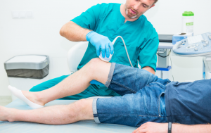
Ultrasound Diagnosis
Beyond x-rays and MRI, the experienced team of knee specialists at USA Pain Center uses ultrasounds for diagnosis. Ultrasounds use sonic waves to capture images of soft tissues and blood vessels, allowing them to detect inflammation in the knee caused by osteoarthritis. Once the source of the inflammation is identified through these images, they can recommend a treatment that can effectively reduce knee pain and swelling, such as genicular artery embolization (GAE).
From Diagnosis to Treatment: Genicular Artery Embolization
Radiologists use X-rays, ultrasounds, and other imaging tools to diagnose knee arthritis, but they cannot treat the condition themselves. An interventional radiologist will use the same tools to diagnose and treat the pain and inflammation.
Pain from knee arthritis often occurs because the inner lining of the joint becomes inflamed. In response, the body sends inflammatory cells to the area, much like it would after an injury. Over time, knee arthritis can lead to the development of new, abnormal blood vessels in the joint lining that also become inflamed. These vessels perpetuate a cycle of inflammation and pain.
An interventional radiologist’s goal is to reduce blood flow to these abnormal vessels through a minimally invasive treatment called genicular artery embolization (GAE). Using ultrasound-guided imaging, an interventional radiologist guides a catheter to the abnormal blood vessels. Tiny particles are injected into those arteries, which will reduce blood flow and lower inflammation.
Benefits of GAE
A main advantage of GAE is that the procedure can be done in an office-based setting. Patients return home shortly after without the need for an extended hospital stay. A recent study published in Osteoarthritis and Cartilage reported minimal risks and side effects from GAE, with only
Researchers note that GAE is over 99 percent successful in reducing knee pain and swelling. Most people reported significant symptom relief after a 12-month follow-up. Additionally, the success of GAE helped delay the need for knee replacement surgery in 5.2 percent of patients with severe knee arthritis. (2)
The team of physicians at USA Pain Center is experienced in performing GAE. Their understanding of the advanced technologies involved helps them carry out the procedure with precision, ensuring patients get effective, long-term relief from knee pain.
Discover if GAE is Right For You

When to Talk to a Specialist About Your Knee Pain
If your knee pain continues for several months or becomes more severe, it’s recommended to see a knee pain specialist. You may also want to consider scheduling a visit if the discomfort leads to joint stiffness that makes daily activities difficult.
Interventional radiologists at USA Pain Center offer genicular artery embolization (GAE) for people with moderate to severe knee arthritis. The procedure is done on an outpatient basis and can be an effective alternative to surgery for those who want to delay knee replacement.
You can easily schedule an appointment online or call the toll-free number to speak with a member of the Care Team. Most health insurance plans are accepted and can be verified once the consultation is booked. Affordable repayment options are available, so you can focus on your treatment without financial stress.
Schedule your consultation today to get back toward improved mobility without pain.
Schedule Your Consultation Online
Sources:
1, 2. Taslakian et al. 2023, Genicular artery embolization for treatment of knee osteoarthritis pain: Systemic review and meta-analysis