
Medically reviewed by
USA Pain Center Medical Review Team
Interventional radiologists focused on knee arthritis
Reviewed: October 2026
An X-ray is typically the first imaging step when a doctor suspects knee arthritis. X-rays show bone changes associated with cartilage loss – but cannot image cartilage itself – making them a useful starting point rather than the complete diagnostic picture.
Knee osteoarthritis develops gradually as cartilage wears down and bone structure changes over time. Understanding what X-rays reveal, where they fall short, and what other imaging may follow can help you make sense of your diagnosis and what it means for the options available to you.
What an X-Ray Shows in a Knee With Arthritis
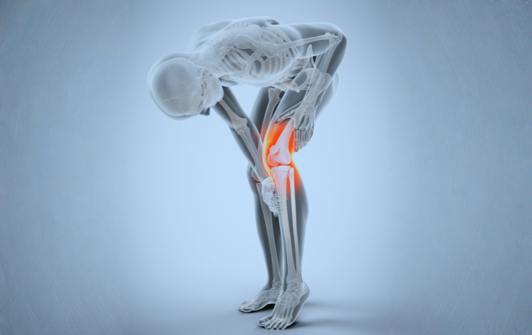
Knee X-rays for arthritis evaluation are taken while you stand – a weight-bearing view – because the load you place on the joint while standing reveals the true degree of joint space narrowing that non-weight-bearing images can underestimate.
A radiologist reviewing a knee X-ray for signs of arthritis will typically look for four findings:
- Joint space narrowing – The gap between the thighbone (femur) and shinbone (tibia) narrows as cartilage wears away. This is the primary indicator of cartilage loss on X-ray.
- Osteophytes (bone spurs) – Bony projections develop at the edges of the joint as the body attempts to stabilize an area of wear.
- Subchondral sclerosis – The bone beneath where cartilage sits becomes denser and more visible on film as arthritis progresses.
- Bone cysts – Small fluid-filled cavities can develop in the bone in more advanced cases.
Together, these findings confirm the presence of osteoarthritis and help your specialist understand how far it has progressed.
The Kellgren-Lawrence Grading Scale
Doctors use a standardized scoring system called the Kellgren-Lawrence (K-L) scale to describe the severity of knee osteoarthritis causes and progression based on what X-ray images reveal. The scale runs from Grade 0 to Grade 4:
- Grade 0: No radiographic signs of arthritis
- Grade 1: Minor bone spur formation, no joint space narrowing
- Grade 2: Definite bone spurs, possible narrowing of joint space
- Grade 3: Moderate joint space narrowing, multiple spurs, bone density changes
- Grade 4: Significant joint space narrowing, large spurs, bone-on-bone contact
The K-L grade plays a role in treatment planning. Patients with moderate arthritis – typically Grade 2 or Grade 3 – are often the most appropriate candidates for minimally invasive interventions. Your K-L grade is one data point, not a prescription. Symptom severity, physical function, and soft tissue findings are also weighed before any treatment path is recommended.
What X-Rays Cannot Show
Cartilage is soft tissue. It does not absorb the electromagnetic energy used in X-ray imaging, so cartilage does not appear on the film. What X-rays reveal is the absence of cartilage – through joint space narrowing where cartilage once existed.
This distinction matters for a few reasons:
Early-Stage Arthritis May Look Normal
Before meaningful joint space narrowing develops, an X-ray can appear nearly normal even when symptoms are already present and cartilage is beginning to wear. This is why early signs of knee arthritis – such as stiffness, aching with activity, and reduced range of motion – are evaluated alongside imaging rather than relying on X-ray alone.
X-Ray Findings May Not Match Your Pain Level
Research confirms that the degree of structural change visible on X-ray does not reliably predict the level of pain or functional limitation a person experiences. Some patients with significant structural findings have manageable symptoms, while others with milder radiographic changes experience significant pain. This is why imaging results are always interpreted in the context of your full clinical picture.
Synovial Inflammation Is Not Visible on X-Ray
Abnormal blood vessel growth in the synovium – the joint lining – is a key contributor to knee pain in osteoarthritis. This type of inflammation, which can be associated with symptoms including knee swelling and persistent aching, is not captured by standard X-ray imaging.
When Other Imaging Is Added
MRI
Magnetic resonance imaging provides detailed views of cartilage, ligaments, and the meniscus that standard X-rays cannot produce. MRI may be ordered when symptoms are more significant than what the X-ray reveals, when another condition may be contributing to knee pain, or when soft tissue involvement needs to be evaluated more precisely before a procedure is considered.
Ultrasound
Ultrasound uses sound waves to visualize soft tissues and blood vessels in and around the knee. It is particularly effective at detecting synovial inflammation – the abnormal overgrowth of blood vessels in the joint lining that is closely linked to pain in knee osteoarthritis. At USA Pain Center, ultrasound is part of the evaluation process used to assess inflammation and identify whether a patient may be a candidate for genicular artery embolization (GAE), a minimally invasive, image-guided procedure that targets the abnormal neovasculature associated with joint inflammation.
What Imaging Means for Your Treatment Options
An X-ray finding does not automatically point toward one treatment. Knee osteoarthritis is managed across a spectrum, and imaging helps specialists understand where on that spectrum a patient falls.
Conservative approaches are typically recommended first, particularly for patients with mild imaging findings and manageable symptoms. Options may include physical therapy to strengthen the muscles that support the knee joint, weight management to reduce load on the joint, anti-inflammatory medications to manage pain and swelling, and activity modification to protect the joint during daily movement.
For patients whose symptoms persist despite conservative care, imaging findings help determine whether more targeted intervention may be appropriate. One option at USA Pain Center is genicular artery embolization (GAE), an outpatient, image-guided procedure that uses imaging to locate and reduce blood flow to abnormal vessels linked to joint inflammation. A review of your X-ray and, in some cases, additional imaging such as ultrasound or MRI is part of the evaluation process used to determine whether GAE is suitable for your situation. Not every patient is a candidate, and a specialist evaluation is the appropriate first step.
A specialist evaluation is the clearest path to an accurate picture of what is happening in your joint and what options may be available based on your specific imaging and symptoms.
Knee Pain Evaluation
Understanding What Your Knee X-Ray Results Mean
If you have been told your X-ray shows signs of knee arthritis, understanding what those findings mean for your options is the right next step. USA Pain Center specialists use advanced imaging and minimally invasive techniques to evaluate knee pain and determine whether options like GAE may be appropriate for your situation.
A consultation includes a review of your imaging, a physical assessment, and a conversation about what your results mean – so you leave with a clear picture of where things stand and what paths may be available.
FAQs About Knee Arthritis X-Rays
- National Institute of Arthritis and Musculoskeletal and Skin Diseases. Osteoarthritis. niams.nih.gov
- Mayo Clinic. Osteoarthritis – Diagnosis and treatment. mayoclinic.org. Updated April 2025.
- Ahmed O, et al. Genicular Artery Embolization for Symptomatic Knee Osteoarthritis: A Comprehensive Review of Techniques and Clinical Outcomes. Cardiovasc Intervent Radiol. 2025 Dec;48(12):1737-1754.
- Lawford BJ, et al. Effects of X-ray-based diagnosis and explanation of knee osteoarthritis on patient beliefs about management: A randomised clinical trial. PLoS Med. 2025;22(2):e1004537.
- Healthline. What to Know About X-Ray for Osteoarthritis of the Knee. healthline.com. Updated April 2024.
Medical disclaimer: This article is for informational purposes only and should not be considered medical advice. Always consult with a qualified healthcare provider for diagnosis, treatment recommendations, and personalized medical guidance.